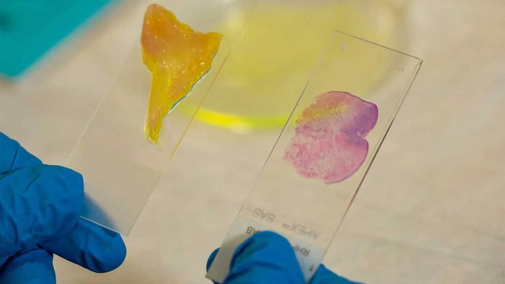
Researchers at Rice University and the MD Anderson Cancer Center of the University of Texas have revealed a new AI-powered microscope. The studies suggest that cancer margins could be tested in minutes by the AI-powered microscope. When surgeons have to remove cancer, the main concern is, did they get it all and to decide whether all the cancer was removed, the margins are examined in depth. The new microscope was designed to analyse large tissue sections rapidly and conveniently, presumably during surgery, to discover the crucial response.
The microscope is designed to view, at cellular resolution, fairly thick pieces of tissue and can allow surgeons to examine tumour margins within minutes of cutting them out. In a study published this week, Rice University engineers and applied physicists developed the telescope and discussed it. The microscope is called the deep learning extended depth-of-field microscope or DeepDOF and utilises deep learning to train a computer algorithm to optimise the variety and post-processing of images.

A standard microscope has a trade-off between spatial resolution and depth-of-field, the researchers say. Which implies only objects at the same distance can be precisely centred from the lens. Details that are also a few millionths of a metre closer or farther from the target of the microscope are fuzzy. Which means the samples from the microscope must be very small and placed on glass slides.
The need to be incredibly thin for the samples means that extracted tissue must be sent to the laboratory where it is frozen or treated with chemicals and cut into slides of razor-thin slices. That’s a time-consuming process that involves advanced machinery. During surgery, it is very unusual for hospitals to have the capacity to make slides for review.
To picture whole pieces of tissue, DeepDOF uses a regular optical microscope combined with an affordable optical facemask that costs less than $10. It can have as much as five times greater depth-of-field than state-of-the-art microscopes today. The facemask is positioned above the objective of the microscope to modulate the light that reaches the microscope. In images taken by the microscope, modulation allows for better control of depth-dependent blur.
The control makes it possible to faithfully retrieve high-frequency texture information over a far wider range of depths than traditional microscopes by deblurring algorithms applied to the captured images. In as little as two minutes, the microscope will capture and process images.

1 Comment
Pingback: Researchers considers Own Immune Cells to Fight Brain Cancer - Craffic